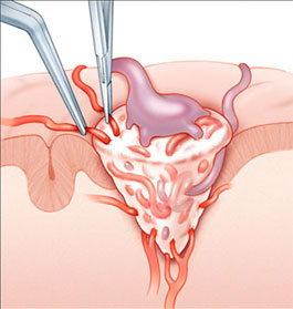


Arteriovenous Malformation (AVM):- arteries carry blood containing oxygen from the heart to the brain, and veins carry blood with less oxygen away from the brain and back to the heart. When an arteriovenous malformation (AVM) occurs, a tangle of blood vessels in the brain or on its surface bypasses normal brain tissue and directly diverts blood from the arteries to the veins. An AVM is composed of an abnormal collection of blood vessels with weakened walls. These abnormal blood vessels have a tendency to bleed. Treatment is recommended to protect against bleeding from the AVM in the future, which may lead, to stroke, permanent disability or even death. About 4 in 100 people with an AVM will have a bleed (haemorrhage) every year. The chances of dying from a bleed is about 10% while the risk of a disability may be as high as 40% with each bleed. The collective risk over a lifetime may be extremely high, especially in a young person. This is why it is very important to treat an AVM.
Surgery has been the traditional method of treating an AVM. The procedure is carried out by a Neurosurgeon who will excise the AVM under general anaesthesia in the operating room. The AVM is adjacent to normal brain but does not contain brain cells within it allowing removal by the surgeon.
Once an AVM is completely taken out surgically, the patient is cured. An AVM does not grow back. The risk of bleeding is thus eliminated immediately after the surgery completely removes the AVM.
Not all AVMs can be treated with surgery. Your surgeon will have ordered tests like the CT scan or MRI, which tell him the size, and exactly where in the brain the AVM is located. The surgeons will then decide if it is safe to remove without serious complications.
The surgical procedure is performed by a highly skilled team of Brain Surgeons, Anaesthetists and the operating room.
Before Surgery- Before the surgery, you will be admitted to the hospital. Routine blood and urine tests and perhaps a chest x-ray and an ECG will have been done as an outpatient to make sure that you are fit for surgery. After midnight, no food or drink is allowed. In some cases, your surgeon might recommend a pre-operative embolization to be performed on the day prior to your scheduled surgery.
You will be taken to the operating room 30 minutes before the operation. After the Anaesthetists put you to sleep, the procedure begins. An area of your head may be shaved. The surgeons will then perform a procedure that is called a craniotomy, which means an opening in the skull. The AVM is then carefully cut out from the surrounding brain. The surgery may take several hours. How long depends on the difficulty encountered by the surgeons. At the end of the surgery, a head dressing will be applied to your head and you will be taken to the Neurosurgical Intensive Care Unit where you will be observed closely.
You will be moved back to your room in 1-2 days. If all goes well, you will be discharged home within a week after surgery. A repeat angiogram will be performed to ensure the AVM has been completely removed.
A period of rest will be required at home. Sometime a course of physiotherapy is required.
The chance of completely curing an AVM using surgical treatment is very high. When completely removed, the AVM will not recur.
Brain AVMs occur in less than 1 percent of the general population. It’s estimated that about one in 200–500 people may have an AVM. AVMs are more common in males than in females.
We don’t know why AVMs occur. Brain AVMs are usually congenital, meaning someone is born with one. But they’re usually not hereditary. People probably don’t inherit an AVM from their parents, and they probably won’t pass one on to their children.
Brain AVMs can occur anywhere within the brain or on its covering. This includes the four major lobes of the front part of the brain (frontal, parietal, temporal, occipital), the back part of the brain (cerebellum), the brainstem, or the ventricles (deep spaces within the brain that produce and circulate the cerebrospinal fluid).